Facilities
University of Rochester/University of Rochester Medical Center:
The University of Rochester, established in 1850, is a private research university located in Rochester, New York. It is known for its rigorous academic environment, fostering interdisciplinary learning and research. The university offers a wide range of undergraduate, graduate, and professional programs across various fields, including arts, sciences, engineering, business, and music. The institution is home to several renowned research centers and institutes, emphasizing innovation and discovery. The University of Rochester is also recognized for its vibrant campus life, promoting a close-knit community and collaborative spirit among students and faculty.
The University of Rochester Medical Center (URMC) is a leading academic medical center that integrates education, research, and patient care. It includes the School of Medicine and Dentistry, the School of Nursing, the Eastman Institute for Oral Health, and a comprehensive healthcare network with numerous hospitals and clinics. URMC is dedicated to advancing medical knowledge and improving healthcare outcomes through cutting-edge research and clinical trials. The center excels in specialties such as cancer treatment, neurology, cardiovascular care, and pediatrics. URMC's commitment to community health and outreach programs further underscores its role in enhancing the well-being of the Rochester community and beyond.
Facility: Mehrmohammadi’s Laboratory
Location: Dr. Mehrmohammadi’s lab is located at Strong Hospital, Rochester, New York
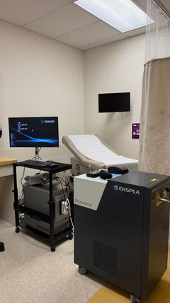
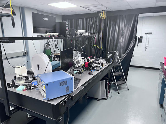
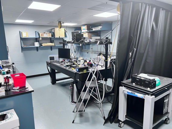
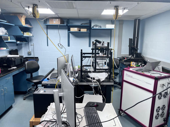
Laboratory Space: State-of-the-art development labs located at the University of Rochester Medical Center (URMC) include two experimental labs (1200 ft2 including a separate 400 ft2 compartment for phantom making area) equipped with optical tables, electronic assembly station, and basic research equipment including digital oscilloscopes, function/arbitrary waveform generators, and multiple system power supplies. Space with seven fully equipped office stations is available for students. A variety of commercial software is available on workstations, including various compilers and computer-aided design packages. MATLAB (MathWorks, Inc.) is a high-performance numeric computation and visualization package integrating numerical analysis, matrix computation, signal processing, and easy-to-use graphics. LabVIEW (National Instruments, Inc.) is a revolutionary single graphical development environment with built-in functionality for data acquisition, instrument control, measurement analysis, and data presentation.
Office Space: Dr. Mehrmohammadi’s research staff and students have 600 ft2 of office space. Each lab member has a personal computer in the office and lab, with connections to the Internet and servers within the University.
Main Equipment:
Ultrasound scanners:
Three fully programmable digital ultrasound systems (Verasonics), one Vantage NXT 256 system with standard frequency and HIFU/arbitrary waveform configurations, Vantage 256, and Vantage 128 high-frequency system with extended burst capability.
Verasonics system is a digital programmable US platform. It provides unique opportunities for research, including full access to transmit and receive procedures and the capability to program them. Most importantly, it provides a unique opportunity to access raw radiofrequency (RF) channel data, which enables us to develop custom-built procedures. Some of the key features of the Verasonics vantage system which is essential for our proposed research, include Ultra-fast imaging using “plane wave” transmission, fast data acquisition ability (up to 14,000 frames/second) into buffer memory, Rapid RF signal data transfer to the host computer (6.6 GB/s), availability of two trigger inputs and one trigger output for synchronizing, and compatibility with various types of transducers. The extended burst module is also enabled on these systems, making it suitable for generating LIFU fields. The systems are also equipped with a universal adaptor module enabling us to interface it to different types of transducers.
Ultrasound transducers:
· 256 and 512 element ring-array transducers: A low-frequency, 256-element ring-array transducer (1.5 MHz) and a mid-frequency, 512-element ring-array transducer (3.5 MHz), each with a 22 cm diameter. These transducers are designed for seamless integration with the Vantage NXT and 256 systems through a 1 to 1 channel adapter, facilitating direct connections.
· Small Animal ring-array transducer: A low-frequency 256-element ring-array transducer (1.5 MHz, HIFU compatible) with an 8 cm diameter. It comes equipped with a 1 to 1 channel adapter for direct connectivity with the Vantage NXT and 256 systems.
· Clinical transducers: Verasonics systems are equipped with three types of probes: a linear array ultrasound transducer (ATL L7-4 x2) operating at 4-7 MHz, a broadband linear array ultrasound transducer (ATL L11-4v) operating at 4-11 MHz, C9-5ICT, a broadband curved array endovaginal ultrasound transducer (ATL C9-5ICT), and a phased-array endoscopic transducer (Siemens AcuNav).
1. High frequency transducers: L35-16 high-frequency transducer for small animal imaging
Lasers and optics:
We have three tunable photoacoustic (nanoseconds) lasers:
1- Ekspla PhotoSonus M+
2- OPOTEK Phocus Mobile system
3- OPOTEK Phocus Core system
4- Quantel Big-sky Nd:YAG pump laser (532 and 1064 nm)
5- Lumics CW, 1470 nm CW laser
6- One Spectra-Physics VGEN-G-20 laser (Single wavelength (green) and high rep rate)
Other optical components include two Ophir power meters, a spectrometer, fiber bundles, optical fiber polishing tools, matching optics including lenses and mirrors, four optical breadboards, fast photodiodes, a CW therapy laser (1470 nm), one vibration-controlled and one regular optical table, and a high-accuracy 3D motorized scanning stage.
Computational tools (computers and software):
The research lab is equipped with seven high-performance computing systems dedicated to data processing and analysis. These include two Intel Xeon 12-core processors at 2.5 GHz with 16 GB RAM, 256 GB SSD, and 1TB HDDs, alongside four Alienware Aurora R13 units boasting 13th Gen Intel Core i9 processors, 64GB RAM, 2TB HDDs, and NVIDIA GeForce GTX 4090 graphics with 32GB GDDR5X memory.
We also utilize a comprehensive suite of software for data and image analysis, including MATLAB, LabView, Field-II Pro, and k-wave for ultrasound simulations; Trace-Pro and Zemax for optical simulations; and AutoCAD, SolidWorks, and 3D Max for mechanical design and simulations.
Other equipment:
Other facilities include an equipped facility for making various types of tissue mimicking phantoms, as well as a fully equipped electronics lab for building necessary peripherals for the system. This includes vacuum oven, hot and steering plates, etc.
1) 3D printer
2) Machine shop facility
3) UV-VIS spectrometer
4) CCD camera for fluorescence imaging.
5) Fiber polishing machine
6) CIRS ultrasound calibration phantom
7) SynDaver tissue-mimicking phantoms
8) Probe Sonicator: Q700 with microtip probe #4418 (1/8”) - Qsonica
9) Rotary evaporator (Rotavapor R-100 – Buchi)
10) Material differential calorimeter
Other Resources at the University of Rochester Medical Center and the University of Rochester
The University of Rochester provides a strong, supportive environment in terms of facilities, mentoring, and physical resources that will enable the proposed project's success. The University of Rochester Medical Center, including the Departments of Imaging Sciences, Surgery, and Microbiology & Immunology, and the School of Medicine and Dentistry, have outstanding research resources to assist investigators and facilitate the progression of their scientific research careers.
The Rochester Center for Biomedical Ultrasound (RCBU): The Rochester Center for Biomedical Ultrasound (RCBU) fosters a distinctive collaborative platform, bringing together experts from engineering, medicine, and applied sciences across the University of Rochester, Rochester General Hospital, and the Rochester Institute of Technology. This initiative paves the way for groundbreaking research in medical diagnostics and therapeutic technologies. RCBU laboratories are advancing the use of ultrasound in diagnosis and discovering new therapeutic applications of ultrasound in medicine and biology. Research at the RCBU spans a wide range of topics in diagnostic and therapeutic ultrasound, including sonoelastography, ultrasound contrast agents including microbubble technology, 3D and 4D ultrasound imaging, volumetric assessment, tissue characterization techniques, lithotripsy, acoustic radiation force imaging, novel therapeutic applications, multi-modal imaging, nonlinear acoustics, and acoustic cavitation.


